Posted on: April 20, 2024
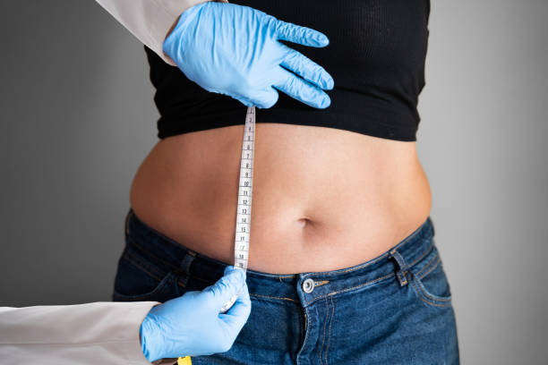
The journey of autologous fat grafting began in the late 19th century. Initially, surgeons experimented with fat transfer for breast reconstruction and facial defects. However, these early attempts often resulted in poor graft survival and significant complications.
Surgeons used simple tools, lacking precision. They faced challenges like resultant scarring and unpredictable outcomes. The understanding of how adipocytes (fat cells) survived post-transfer was minimal.
By the mid-20th century, advancements in medical technology revolutionized fat grafting. The introduction of the cannula, a thin tube used to transfer fat, allowed plastic surgeons to perform the procedure with more accuracy.
This period saw a pivotal shift towards understanding the importance of blood supply for graft survival. Researchers discovered that ensuring a rich blood supply to the transferred fat significantly improved its survival rate.
In recent years, the field has seen remarkable improvements. Today’s plastic surgeons employ highly sophisticated techniques that enhance both safety and effectiveness.
Techniques now focus on minimizing damage to adipocytes during transfer. Surgeons meticulously ensure that fat is deposited in areas with ample blood vessels to support survival. This attention to detail has led to higher success rates and fewer complications.
Modern procedures also benefit from better pre-operative planning and post-operative care, reducing risks associated with graft necrosis.
Following the evolution of autologous fat grafting, it’s crucial to understand how fat necrosis develops. The process begins when adipocytes, or fat cells, suffer damage during the transfer. This damage leads to ischemia, a condition where blood supply to the adipose tissue is insufficient.
Ischemia triggers a cascade of inflammatory responses. Inflammatory cells, including macrophages, rush to the site. They release inflammatory factors that further aggravate the condition.
The presence of these inflammatory cells marks the start of an intense inflammatory reaction. This reaction is not just a defense mechanism but also a contributor to cell death. As inflammation progresses, it disrupts the lipid structure within fat cells.
This disruption can lead to infection, worsening the situation. The area becomes a battleground for inflammatory reactions and attempts at healing.
Over time, if unresolved, this inflammation leads to fat necrosis. Dead and damaged adipocytes accumulate, forming necrotic tissue. This tissue lacks any viable blood supply and can become a long-term complication if not addressed.
Risk factors increasing this outcome include poor technique during fat transfer, existing health conditions that impair healing, and inadequate post-procedure care.
Fat grafting, especially in the face, demands precise techniques to ensure fat cell survival. Hypoxia occurs when these cells don’t get enough oxygen. This lack of oxygen can kill fat cells, leading to complications like necrosis.
After a transfer, the relocated fat struggles to establish a new blood supply. Oxygen tension is critical during this period. Without adequate oxygen, cells begin to die, compromising the graft’s success.
The link between hypoxia and fat necrosis is significant. When transferred fat doesn’t receive sufficient oxygen, it increases the risk of necrosis. This condition not only affects cosmetic outcomes but can also lead to further complications requiring corrective procedures.
Understanding this relationship helps surgeons anticipate and mitigate risks associated with facial fat grafting.
To combat hypoxia and improve outcomes, several strategies are crucial. First, ensuring optimal oxygen supply during and after the procedure is key. Techniques that enhance blood flow to the transplanted fat can significantly reduce hypoxia-related risks.
Hyperbaric oxygen therapy (HBOT) has emerged as a promising adjunctive treatment. By increasing oxygen tension around the graft site, HBOT can promote better healing and cell survival.
Adipocyte browning involves the conversion of white fat cells into beige fat cells. This process can affect facial fat grafting outcomes. White fat stores energy, while beige fat burns it, producing heat. Factors triggering this conversion include cold exposure and certain inflammatory responses.
Browning reduces the volume of transferred fat, potentially leading to graft necrosis. It’s crucial for cosmetic surgeons to understand how adipocyte browning impacts graft survival.
The inflammatory response plays a significant role in adipocyte browning. Post-surgical inflammation might accelerate this process. It could lead to an undesirable outcome in facial aesthetics by reducing the longevity of the fat graft.
Current research focuses on minimizing inflammation to prevent or slow down the browning of transplanted fat cells. This is vital for improving fat graft survival and achieving desired aesthetic results.
Scientists are exploring ways to prevent or reverse adipose tissue browning. Their goal is to enhance the longevity and stability of facial fat transfers. Understanding the underlying mechanisms of browning is key to developing effective strategies.
Fibrosis plays a critical role in the aftermath of fat transfer necrosis, particularly when fat cells undergo trauma. This condition often leads to hardening of the fatty tissue, making it firm and less pliable. Such changes can severely impact facial contouring outcomes, as the transferred fat fails to integrate smoothly with the surrounding tissues.
After a fat transfer procedure, some fat cells may not survive the process, leading to necrosis. The body responds by initiating an inflammatory reaction to remove dead cells. Over time, this area can develop fibrosis, characterized by the formation of scar-like tissue. This scarring can cause irregularities and firmness in the treated areas, detracting from the smooth, natural appearance desired.
Addressing fibrosis requires targeted therapeutic strategies aimed at reducing scarring and promoting healthier tissue regeneration. Treatments may include:
Researchers have explored various models, including studies on nude mice, to understand how to best enhance healing post-fat transfer and minimize complications like fibrosis. These studies suggest that improving blood supply to the transferred fat can reduce the risk of necrosis and subsequent fibrosis.
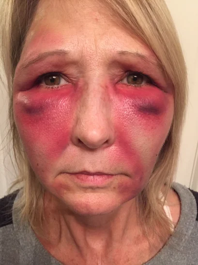
Early detection of fat necrosis after facial fat grafting is crucial. Diagnostic imaging can reveal areas with poor graft viability. Ultrasound and MRI are effective in identifying oil cysts and areas of necrosis. These methods allow for a targeted approach in managing complications.
Patients may notice periorbital swelling or firm nodules as early signs. It’s essential to report these symptoms promptly. This enables swift diagnostic measures, reducing the risk of long-term damage.
Managing fat necrosis involves both conservative measures and surgical interventions. Initially, conservative approaches like massage and warm compresses can help resolve minor cases. They reduce pressure in affected areas, promoting healing.
For more severe instances, surgical removal of necrotic tissue might be necessary. This ensures healthy tissue regeneration. Incorporating m2 macrophages into the treatment can aid in recovery by enhancing tissue repair mechanisms.
Educating patients on the potential risks and postoperative care is vital. They should understand the importance of immediate reporting of any unusual symptoms post-surgery. Regular follow-up appointments are critical for monitoring recovery and detecting any signs of necrosis early.
Proper patient education minimizes the risk of complications and ensures a smoother recovery process. It empowers patients to take an active role in their healing journey.
Stem cell therapy has emerged as a cutting-edge solution for treating fat necrosis. This method employs the body’s natural healing mechanisms to repair damaged tissues. Researchers are exploring how stem cells can enhance the survival rate of fat grafts.
They believe these cells can regenerate lost or damaged tissue, thus reducing the risk of necrosis. Early results have shown promise, with some patients experiencing improved outcomes and less complication post-procedure.
Regenerative medicine techniques are also making strides in addressing fat necrosis. These approaches focus on restoring the function of fat tissue rather than just removing or replacing it.
By using growth factors, cytokines, and peptides, scientists aim to create a more conducive environment for fat grafts to thrive. This method not only helps in preventing necrosis but also promotes long-term graft retention and integration.
The development of novel pharmacological agents is another area of interest. These drugs aim to prevent the occurrence of fat necrosis or mitigate its severity by improving blood supply to the transplanted fat.
Ongoing research is dedicated to identifying compounds that can increase angiogenesis (the formation of new blood vessels) within the grafted fat. Such advancements could revolutionize how we approach facial fat transfer procedures, making them safer and more reliable.
Autologous fat grafting has evolved, offering a promising solution for facial rejuvenation, yet it’s not without its challenges. Understanding the pathogenesis of fat necrosis, from hypoxia to fibrosis, is crucial in managing and preventing this complication. Innovations in treatment have significantly improved outcomes, but awareness and early intervention remain key. Your knowledge and proactive approach can make a difference in experiencing the benefits of facial fat transfer while minimizing risks. Always consult with a skilled professional who stays abreast of the latest advancements in the field.
Taking charge of your aesthetic journey means staying informed about potential complications like fat necrosis and knowing the cutting-edge treatments available. If you’re considering facial fat transfer, arm yourself with information and choose a provider who prioritizes safety and efficacy. Let’s embrace innovation responsibly, ensuring beauty enhancements come with minimized risks. Ready to take the next step? Consult a trusted expert today.
Facial fat transfer necrosis occurs when transferred fat cells fail to re-establish blood supply, leading to their death and potential complications. It’s a rare but serious condition that can affect the outcome of autologous fat grafting procedures.
Hypoxia, or lack of oxygen, plays a critical role in fat transfer complications by preventing the newly transferred fat cells from surviving. This can lead to cell death and increase the risk of necrosis in the treated area.
The browning mechanism refers to the process where white adipose tissue (fat) turns into brown-like adipose tissue under certain conditions, potentially impacting the viability and appearance of transferred facial fat.
Yes, fibrosis, which is the formation of excess fibrous connective tissue, can occur as a response to necrosis following a facial fat transfer. It may lead to hardening or thickening in the treatment area.
Managing facial fat necrosis involves timely intervention with treatments like massage, use of compression garments, or more invasive procedures such as debridement or revision surgery, depending on severity.
Yes, recent innovations in treating fat necrosis include advanced surgical techniques for removal and repair, improved methods for ensuring the viability of transplanted fat, and novel therapies aimed at reducing inflammation and promoting healing.
Understanding the pathogenesis of fat necrosis is crucial for preventing this complication during autologous fat grafting. It enables clinicians to optimize techniques for harvesting and transferring fat, thereby improving patient outcomes.